Epilepsy after Concussion – Seizure
Hippocampal Model of ESD (Epilepsy after Concussion)
We propose a neurophysiological model of hippocampal-limbic and temporal lobe dysfunction which can explain the neurobehavioral symptoms characteristic of epilepsy spectrum disorder (epilepsy after concussion). The temporal lobe both in its limbic and isocortical components is responsible for the majority of ictal behavioral changes resulting from epilepsy after concussion seizure discharge (Gloor 1991). Within the temporal lobe, the hippocampus contains pyramidal cells which have multiple excitatory connections. These in turn are controlled by the inhibitory influence of granule cells in a ratio of about 200 to 1. Thus, damage to granule cells, can result in disinhibition of the excitatory activity of the hippocampal pyramidal cells. Although the hippocampal pyramidal cells remain healthy, they lack the inhibitory controls exercised on them by granule cells and are essentially allowed to discharge unchecked.
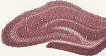
Diagram of the Hippocampus
Cellular physiology – The hippocampus has been thoroughly studied and often is associated with local epileptiform activity. Cajal divided the hippocampus into seven layers (Gloor 1991). The two most relevant to our discussion are the pyramidal layer (Ammon’s horn) and the stratum moleculare which includes the dentate gyrus. The hippocampal pyramidal cells (HPCs) provide the excitatory output of the hippocampus. The HPCs are organized in stacked, linear, geometric arrays, each cell with the same longitudinal orientation. This arrangement allows the HPCs to act as a battery (Foster et al. 1988). In some models, only five cells are required to initiate synchronous firing in up to 20 cells, which in turn potentiate larger groups of cells (Miles & Wong 1983). This effect is even more critical since HPCs are capable of spontaneous discharges and have extensive connections with other cortical and subcortical regions. Thus, the misfiring of a single cell can have influence on many other HPCs and subsequently can effect a greater collection of brain cells further downstream.
The excitatory output of the HPCs is modulated via inhibitory granule cell neurons arising from the dentate gyrus (Isaacson & Pribram 1975, Prince 1983). Because hippocampal granule cells inhibit HPCs in a ratio of approximately 1 to 200, damage to only a few hippocampal inhibitory cells can exert a disproportionate effect on the regulation of HPC output (Foster et al. 1988). Therefore, the death or malfunction of only a few granule cells can cause hyperactivity in many times more HPCs.
Disproportionate inhibitory effect of hippocampal granule cells on the HPCs is even more critical given other aspects of hippocampal circuitry. Incoming afferent input from entorhinal cortex and the fimbria provide an excitatory stimulus to the HPCs. Inhibitory granule cells inhibit HPCs both directly and via a series of feed-forward circuits which retard the incoming afferent input from the fimbria and entorhinal cortex (Prince 1983, Oliver & Miller 1985). Damage to the inhibitory granule cells can reduce feed-forward inhibition of the excitatory input as well as reduce the inhibitory control of excitatory HPC output.
This basic effect is further amplified by other aspects of the hippocampal circuitry. The excitatory output of HPC feedback to inhibitory granule cells also has GABA mediated inhibitory feedback on pyramidal cells. Injury to the local GABA mediated inhibitory circuitry unmasks additional HPC excitatory interneurons circuitry. This unmasking can produce bursting (i.e., sudden synchronous abnormal spontaneous electrical discharges) from populations or neurons (Oliver & Miller 1985). GABA blockade can also produce this effect.
Hippocampal Dynamics